AS Biology, by topic.
Cells to immunity, written for the paper in front of you: definitions, mechanisms, calculations and the usual exam traps.
1Cell Structure
Microscopy and measurement
Magnification describes how many times larger an image is than the specimen; resolution is the minimum distance at which two points can still be distinguished. A larger image is not automatically more informative if resolution has not improved.
Convert every value to the same unit before calculating. A calibrated eyepiece graticule changes value whenever the objective lens changes. A stage micrometer supplies the known scale used to calibrate it.
Preparing and viewing specimens
A light microscope forms an image when visible light passes through, or is reflected from, a specimen and is focused by glass lenses. A thin specimen is needed so that sufficient light is transmitted. Temporary wet mounts are suitable for living material; a small sample is placed in a drop of water and covered with a coverslip lowered at an angle to reduce trapped air bubbles. A squash spreads soft tissue into a thin layer, while a smear is useful for liquids such as blood or microbial cultures. Permanent sections are normally fixed, dehydrated, embedded, sliced with a microtome and stained.
Many cell components have little natural contrast. Stains absorb differently in different structures, making boundaries easier to distinguish. Iodine can make starch-containing material visible, methylene blue can increase nuclear contrast, and differential stains can distinguish tissues or groups of bacteria. A stain changes the appearance of the specimen, so colour in a prepared slide is not automatically the cell's natural colour.
Calibrating an eyepiece graticule
An eyepiece graticule is an arbitrary scale in the eyepiece. Its divisions have no fixed length until they are compared with the known scale on a stage micrometer. Superimpose both scales, align two marks, and find the known stage-micrometer distance that corresponds to a counted number of eyepiece divisions.
Repeat the calibration whenever the objective lens is changed. A higher objective magnification makes each eyepiece division represent a smaller actual distance. Record enough significant figures during the calculation, but round the final measurement to match the precision of the scale.
Resolution and microscope choice
| Microscope | Useful features | Limitations and image |
|---|---|---|
| Light microscope | Can show living specimens, movement and natural or stained colour; relatively simple preparation | Resolution is about 200 nm, so membranes and ribosomes cannot be resolved; image is two-dimensional |
| Transmission electron microscope (TEM) | Electrons pass through an ultrathin section; very high resolution reveals internal ultrastructure | Specimen must be dead and viewed in a vacuum; preparation may create artefacts; image is a two-dimensional section |
| Scanning electron microscope (SEM) | A beam scans the surface and detected electrons reveal surface topography | Resolution is lower than TEM; specimen is dead; the image has a three-dimensional appearance but is displayed on a flat surface |
Electron beams have a much shorter effective wavelength than visible light, so electron microscopes can distinguish points that are much closer together. Resolution, not advertised magnification, determines whether extra detail is genuine. False colour may be added to electron micrographs after imaging and does not represent the original specimen colour.
Cell ultrastructure
Eukaryotic cells contain membrane-bound organelles. The nucleus stores DNA; rough endoplasmic reticulum and ribosomes synthesise proteins; the Golgi modifies and packages them; mitochondria produce ATP; lysosomes contain hydrolytic enzymes. Chloroplasts, cellulose walls and a large permanent vacuole distinguish typical plant cells.
Prokaryotes are smaller and lack a nucleus and membrane-bound organelles. Their circular DNA lies free in the cytoplasm, plasmids may carry additional genes, 70S ribosomes synthesise proteins, and a peptidoglycan wall surrounds the membrane.
| Structure | Recognition feature | Function link |
|---|---|---|
| Cell surface membrane | About 7-10 nm wide; appears as two dark lines in a TEM | Controls exchange, receives signals and supports cell recognition |
| Nucleus and nucleolus | Double nuclear envelope with pores; dense nucleolus; chromatin in nucleoplasm | DNA controls transcription; the nucleolus makes rRNA and assembles ribosomal subunits |
| Rough endoplasmic reticulum | Flattened membrane sacs with attached ribosomes | Synthesises, folds and transports proteins for secretion or membranes |
| Smooth endoplasmic reticulum | Tubular membranes without ribosomes | Synthesises lipids and carries out detoxification |
| Golgi body | Stacked flattened cisternae with budding vesicles | Modifies proteins, sorts them and packages them into vesicles; forms lysosomes |
| Mitochondrion | Double membrane; inner membrane folded into cristae; matrix within | Cristae hold electron carriers and ATP synthase; the matrix contains enzymes for aerobic respiration |
| Chloroplast | Double envelope; thylakoids stacked as grana; fluid stroma; starch grains may be present | Thylakoid membranes carry out light-dependent reactions; the stroma contains Calvin-cycle enzymes |
| Ribosome | Small dense granule; 80S in eukaryotic cytoplasm and 70S in prokaryotes, mitochondria and chloroplasts | Site of translation, where amino acids are joined into a polypeptide |
| Lysosome | Small single-membrane vesicle containing hydrolytic enzymes | Digests material taken into the cell and breaks down worn organelles |
| Centriole | Cylinder of microtubules, usually found as a pair near an animal-cell nucleus | Organises spindle microtubules during nuclear division |
| Cellulose cell wall | Rigid layer outside the plant cell surface membrane; crossed by plasmodesmata | Resists osmotic expansion, supports the cell and provides pathways between adjacent cells |
| Large permanent vacuole | Fluid-filled compartment bounded by a tonoplast | Stores solutes and pigments and helps maintain turgor |
From organelles to secreted protein
A gene in the nucleus is transcribed and the mRNA leaves through a nuclear pore. A ribosome on rough endoplasmic reticulum translates the mRNA, and the growing protein enters the RER lumen for folding. Transport vesicles carry it to the cis face of the Golgi body. As it passes through Golgi cisternae it may be modified, for example by addition of carbohydrate. A secretory vesicle buds from the trans face, moves to the cell surface membrane and fuses with it, releasing the product by exocytosis.
ATP as the cell's immediate energy currency
ATP consists of adenine, ribose and three phosphate groups. Hydrolysis of the terminal phosphate forms ADP and inorganic phosphate and releases a small, manageable quantity of energy. ATP can also phosphorylate another molecule, making it more reactive. Because ATP is rapidly regenerated from ADP and phosphate, it couples energy-releasing reactions such as respiration to energy-requiring processes including active transport, biosynthesis, movement and vesicle trafficking. ATP is an immediate energy carrier, not a long-term energy store.
Prokaryotic and eukaryotic cells
| Feature | Prokaryotic cell | Eukaryotic cell |
|---|---|---|
| Typical size | Usually about 0.5-5 µm | Often about 10-100 µm |
| Genetic material | One circular, naked DNA molecule in a nucleoid; plasmids may occur | Linear DNA associated with histones inside a nucleus |
| Internal membranes | No membrane-bound organelles | Membrane-bound organelles compartmentalise reactions |
| Ribosomes | 70S | 80S in cytoplasm; 70S inside mitochondria and chloroplasts |
| Cell wall | Peptidoglycan in bacteria | Cellulose in plants, chitin in fungi, absent from animals |
| Cell division | Binary fission | Mitosis or meiosis |
A typical bacterium may also possess a slime capsule for protection and adhesion, pili for attachment or DNA transfer, and one or more flagella for movement. Its cell surface membrane carries out some metabolic processes that occur on organelle membranes in eukaryotes. Do not call bacterial DNA a chromosome inside a nucleus: it is located in the nucleoid region and is not surrounded by a nuclear envelope.
Viruses are acellular particles
A virus is not a cell. It contains genetic material, either DNA or RNA, enclosed by a protein capsid. Some viruses also have a phospholipid envelope derived from a host membrane and bearing attachment proteins. Viruses have no cytoplasm, ribosomes or independent metabolism, so they cannot synthesise ATP or proteins by themselves.
Replication occurs only inside a living host cell. Viral attachment proteins bind to complementary receptors, the viral nucleic acid enters the cell, and the host's enzymes and ribosomes are redirected to copy viral genetic material and make capsid proteins. New particles assemble and leave by cell lysis or budding. Their receptor specificity helps explain why a virus infects particular cell types or host species.
Drawing and comparison
A plan diagram shows the distribution of tissues, not individual cells. Use single clear lines, no shading, a ruled label line, a title and a magnification or scale bar. For a comparison, pair each difference in the same sentence or table row.
In a high-power cell drawing, draw only boundaries that can actually be seen and keep their relative proportions. In a low-power plan diagram, show the outlines and relative thicknesses of tissue layers without filling them with invented cells. A photomicrograph is produced using a light microscope; an electron micrograph is produced using an electron microscope. Always inspect the scale bar before inferring size.
Active recall checkpoint
2Biological Molecules
Monomers, polymers and water
Condensation joins monomers and releases water; hydrolysis adds water to break a covalent bond. Monosaccharides form glycosidic bonds, amino acids form peptide bonds, and glycerol plus fatty acids form ester bonds.
Water is polar, so it is a solvent for ions and other polar molecules. Hydrogen bonding produces cohesion, a high specific heat capacity and a high latent heat of vaporisation. These properties support transport, temperature stability and evaporative cooling.
Carbohydrates and glycosidic bonds
Monosaccharides are single sugar units. Glucose is a hexose with the formula \(\mathrm{C_6H_{12}O_6}\). In aqueous solution it usually forms a ring. Alpha-glucose and beta-glucose are isomers: they have the same molecular formula but differ in the position of the hydroxyl group on carbon 1. This small structural difference produces polysaccharides with very different shapes and functions.
A condensation reaction between two monosaccharides forms a glycosidic bond and releases water. Two alpha-glucose molecules can form maltose; glucose and fructose form sucrose; glucose and galactose form lactose. The reverse reaction is hydrolysis, in which water is added to split the glycosidic bond.
Starch, glycogen and cellulose
| Polysaccharide | Structure | How structure suits function |
|---|---|---|
| Amylose | Unbranched alpha-glucose chain with \(\alpha\)-1,4 bonds; coils into a helix | Compact and insoluble, so it stores glucose without lowering water potential |
| Amylopectin | Alpha-glucose with \(\alpha\)-1,4 chains and \(\alpha\)-1,6 branch points | Many terminal ends allow enzymes to release glucose rapidly |
| Glycogen | Similar to amylopectin but more highly branched | Compact animal and fungal store; many ends support rapid hydrolysis during high demand |
| Cellulose | Unbranched beta-glucose chains with \(\beta\)-1,4 bonds; alternate monomers are inverted | Straight parallel chains form many hydrogen bonds, producing strong microfibrils in plant cell walls |
Starch is a mixture of amylose and amylopectin. These storage molecules are large and insoluble, so they do not diffuse from cells and do not create an osmotic influx of water. Cellulose is not a storage polymer: microfibrils cross-link into fibres that have high tensile strength, resist turgor pressure and prevent plant cells from bursting.
Triglycerides and phospholipids
A triglyceride forms when glycerol reacts with three fatty acids. Three condensation reactions create three ester bonds and release three water molecules. Saturated fatty acids have no carbon-carbon double bonds; their straight hydrocarbon chains pack closely. Unsaturated fatty acids contain one or more carbon-carbon double bonds, which introduce bends and usually lower the melting point.
Triglycerides contain a high proportion of energy-rich carbon-hydrogen bonds, so oxidation releases more energy per gram than carbohydrate. They are non-polar and insoluble, allowing energy storage without affecting cell water potential. Fat beneath the skin can insulate, and adipose tissue can cushion organs. Oxidation also produces metabolic water.
A phospholipid contains glycerol, two fatty acids and a phosphate-containing group. Its phosphate head is polar and hydrophilic, while the fatty-acid tails are non-polar and hydrophobic. This amphipathic character causes phospholipids to form bilayers in water, with heads facing aqueous environments and tails sheltered inside.
Structure determines function
Compact, insoluble glucose stores. Amylopectin and glycogen are branched, providing many ends for rapid hydrolysis.
Parallel beta-glucose chains form hydrogen-bonded microfibrils with high tensile strength.
Energy-dense and insoluble; oxidation yields metabolic water. Phospholipids are amphipathic and form bilayers.
Primary sequence controls folding into secondary, tertiary and sometimes quaternary structure.
Globular proteins are compact and usually soluble, making them suitable as enzymes, hormones and transport proteins. Fibrous proteins have repeated sequences and extensive cross-linking, providing structural strength.
Amino acids and peptide bonds
Every amino acid has a central carbon bonded to an amino group, a carboxyl group, a hydrogen atom and a variable R group. The R group gives each amino acid its chemical properties. A condensation reaction between the carboxyl group of one amino acid and the amino group of another forms a peptide bond. A dipeptide contains two amino-acid residues; many residues joined together form a polypeptide.
The order of amino acids is the primary structure. Because R groups interact differently with water and with one another, the primary sequence determines how the polypeptide folds and therefore its biological function. A change to the amino-acid sequence can alter bonding, shape, stability or the properties of a binding site.
The four levels of protein structure
| Level | Description and stabilising interactions |
|---|---|
| Primary | The amino-acid sequence, held by covalent peptide bonds |
| Secondary | Regular alpha-helices or beta-pleated sheets formed by hydrogen bonds between peptide groups |
| Tertiary | The overall three-dimensional shape of one polypeptide, stabilised by hydrogen bonds, ionic attractions, disulfide bonds and hydrophobic interactions between R groups |
| Quaternary | The association of two or more polypeptide subunits, sometimes with a non-protein prosthetic group |
High temperature or an extreme pH can disrupt the interactions that maintain secondary, tertiary and quaternary structure. The primary structure usually remains intact because peptide bonds are not broken under ordinary denaturing conditions. Loss of the precise three-dimensional shape can make a protein non-functional.
Globular and fibrous proteins
Globular proteins fold into compact shapes, often with hydrophilic R groups facing water and hydrophobic R groups sheltered inside. This commonly makes them soluble and suitable for dynamic roles such as enzymes, antibodies, membrane transport and oxygen carriage. Fibrous proteins have long, repeated structures and are usually insoluble, making them suitable for strength and support.
Haemoglobin is a globular conjugated protein with four polypeptide subunits. Each subunit contains a haem prosthetic group with an \(\mathrm{Fe^{2+}}\) ion that can bind one oxygen molecule reversibly, so one haemoglobin molecule can carry four oxygen molecules. Its quaternary structure allows cooperative binding: attachment of one oxygen changes the protein's shape and increases the affinity of the remaining subunits.
Collagen is fibrous. Three polypeptide chains wind together into a triple helix, with glycine occurring frequently so the chains can pack closely. Covalent cross-links form between molecules, and many molecules assemble into fibrils and fibres. This hierarchy gives high tensile strength in tendons, skin, cartilage and the walls of blood vessels.
Why water is biologically important
Oxygen attracts the shared electrons in each O-H bond more strongly than hydrogen, making water polar. The slightly positive hydrogen of one molecule is attracted to the slightly negative oxygen of another, forming a hydrogen bond. Each bond is weak, but very large numbers together produce important emergent properties.
| Property | Biological consequence |
|---|---|
| Excellent solvent for ions and polar molecules | Provides the medium for metabolic reactions and transports solutes in blood, tissue fluid and plant sap |
| High specific heat capacity | Much energy is needed to change temperature, buffering cells and aquatic habitats against rapid fluctuation |
| High latent heat of vaporisation | Evaporation removes substantial energy, enabling cooling by sweating and transpiration |
| Cohesion and surface tension | Hydrogen bonding maintains continuous water columns in xylem and supports small organisms at the surface |
| Water is a reactant | Hydrolysis splits biological molecules; water is also used in photosynthesis |
| Ice is less dense than liquid water | Ice floats and insulates water below, allowing aquatic life to survive cold conditions |
Biochemical tests
| Substance | Procedure | Positive result |
|---|---|---|
| Reducing sugar | Add Benedict reagent and heat in a water bath | Blue to green/yellow/orange/brick-red precipitate |
| Non-reducing sugar | Boil with dilute acid, neutralise, then Benedict test | Coloured precipitate |
| Starch | Add iodine in potassium iodide | Blue-black |
| Protein | Biuret reagent | Lilac/purple |
| Lipid | Ethanol emulsion test, then add water | White emulsion |
For the non-reducing-sugar test, first test a fresh portion with Benedict reagent. A negative initial result is essential: otherwise a later precipitate could have come from reducing sugar already present. Heat another portion with dilute hydrochloric acid to hydrolyse non-reducing sugar, cool it, neutralise the acid with sodium hydrogencarbonate or sodium hydroxide, then repeat the Benedict test.
In the emulsion test, shake the sample thoroughly with ethanol so any lipid dissolves, then pour the ethanol extract into water. Lipid forms tiny suspended droplets that scatter light, producing a cloudy white emulsion. Water alone is not a valid lipid solvent, and heating is not required.
A colorimeter measures how much light is transmitted or absorbed at a selected wavelength. Zero the instrument with an appropriate blank, use clean cuvettes in the same orientation and remove fingerprints. A calibration curve should place known concentration on the x-axis and absorbance on the y-axis. If an unknown lies outside the calibrated range, dilute it by a known factor, repeat the measurement and correct the final concentration for the dilution.
Active recall checkpoint
3Enzymes
How enzymes work
An enzyme is a globular protein with a specific active site. Substrate binding forms an enzyme-substrate complex and lowers activation energy by orientating reactants, straining bonds or creating a favourable microenvironment. In the induced-fit model, binding changes the active-site shape to improve complementarity.
Temperature and pH alter rate by changing kinetic energy, collision frequency and the bonds that maintain tertiary structure. Denaturation changes active-site shape; it is not the same as the enzyme being killed.
Specificity, activation energy and induced fit
The active site is a three-dimensional region formed when the polypeptide folds. Amino-acid R groups within it create a particular shape and chemical environment. Only a substrate with sufficiently complementary shape and charge can bind and form an enzyme-substrate complex, which explains enzyme specificity.
The lock-and-key model treats the active site as rigid. The induced-fit model is more complete: initial contact with the substrate changes the enzyme's conformation, bringing catalytic groups into better positions and placing strain on substrate bonds. The enzyme provides an alternative reaction pathway with a lower activation energy. It does not change the overall energy released, the reaction's equilibrium or the nature of the products.
After catalysis, the products have a different shape or charge distribution and leave the active site. The enzyme is chemically unchanged and can catalyse another reaction. Enzymes increase both forward and reverse reaction rates; the direction of net change depends on substrate and product conditions.
Measuring enzyme activity
Activity can be followed by measuring the disappearance of substrate or appearance of product. Suitable measurements include gas volume, mass loss, pH change, colour or absorbance, or the time taken for a visible endpoint. A continuous method produces many readings from one reaction mixture; a discontinuous method removes samples at intervals or uses separate tubes stopped at different times.
Rate must be distinguished from time. If the same fixed endpoint is used, a shorter time means a faster reaction and relative rate can be calculated as \(1/t\). Where a graph of product against time is available, rate is the gradient. The initial tangent gives the least-confounded estimate because substrate has barely been depleted and little product has accumulated.
Rate and inhibition
At low substrate concentration, rate rises because successful collisions become more frequent. At high concentration, active sites are saturated and the rate approaches \(V_{\max}\). Increasing enzyme concentration raises \(V_{\max}\) if substrate is not limiting.
Temperature
As temperature rises, enzyme and substrate molecules gain kinetic energy, collide more frequently and form more enzyme-substrate complexes per second. Rate therefore rises towards an optimum. Above the optimum, increased vibration disrupts hydrogen bonds and ionic interactions that maintain tertiary structure. The active site loses complementarity, fewer complexes form and rate falls steeply. A low temperature normally causes reversible slowing; high-temperature denaturation is often irreversible.
pH
Changing hydrogen-ion concentration changes the charges on amino-acid R groups. This can disrupt ionic and hydrogen bonds that maintain the active site's shape, or alter the charge of groups directly involved in substrate binding and catalysis. Each enzyme has an optimum pH related to its normal environment. A pH-rate curve is often roughly bell-shaped, but its exact form depends on the enzyme.
Enzyme and substrate concentration
With excess substrate, doubling enzyme concentration approximately doubles the initial rate because twice as many active sites are available. If substrate becomes limiting, this proportionality is lost. With fixed enzyme concentration, raising substrate concentration initially increases rate, but the curve levels as an increasing fraction of active sites are occupied. At saturation, almost every active site is continually processing substrate and the rate is close to \(V_{\max}\).
The Michaelis-Menten constant, \(K_m\), is the substrate concentration at which rate is half \(V_{\max}\). Under the simple model, a smaller \(K_m\) means half-maximal rate is reached at a lower substrate concentration and therefore indicates greater enzyme-substrate affinity. Read \(V_{\max}\) from the plateau first, halve it, then project from that rate to the substrate-concentration axis to estimate \(K_m\).
Enzyme inhibition
| Inhibitor | Binding and mechanism | Effect on rate curve |
|---|---|---|
| Competitive | Has enough similarity to the substrate to occupy the active site; reduces the chance of substrate binding | Its effect can be reduced by high substrate concentration; \(V_{\max}\) can still be reached, but apparent \(K_m\) increases |
| Non-competitive | Binds at an allosteric site and changes enzyme conformation, so fewer active sites function effectively | Extra substrate cannot restore the original \(V_{\max}\); in the pure model \(K_m\) is unchanged |
| Irreversible | Forms a permanent association, often a covalent bond, or permanently disrupts the active site | Reduces the effective enzyme concentration until new enzyme is synthesised |
An inhibitor's effect depends on its concentration. More inhibitor molecules create more frequent encounters with enzyme molecules. For a competitive inhibitor, both inhibitor and substrate concentrations determine the proportion of active sites occupied by each. Never identify inhibition type from a single rate value; compare how the curve responds across a range of substrate concentrations.
Measuring initial rate
Measure product formed or substrate lost over time, then draw a tangent at time zero. Initial rate avoids the later effects of substrate depletion, product accumulation and reverse reaction.
Planning a valid enzyme investigation
Change only the independent variable and specify how it is produced: use thermostatically controlled water baths for temperature, buffer solutions for pH, or serial dilutions for concentration. Control enzyme and substrate volumes and concentrations, total reaction volume, mixing, sampling interval and the time between combining reagents and taking the first reading. Equilibrate solutions to the chosen temperature before mixing.
Use at least five appropriately spaced values, including values on both sides of any expected optimum. Repeat each condition and calculate a mean; investigate anomalous results instead of deleting them without reason. Plot the independent variable with units on the x-axis and initial rate with units on the y-axis. A smooth biological trend is normally more appropriate than joining every point with straight segments.
Immobilised enzymes
An immobilised enzyme is held in place rather than freely mixed with the reaction solution. One school-laboratory method mixes enzyme with sodium alginate and releases drops into calcium chloride. Calcium alginate beads form and trap the enzyme; small substrate and product molecules diffuse through the gel.
| Advantage | Reason |
|---|---|
| Product is not contaminated with enzyme | Downstream purification is easier and the enzyme is retained in the reactor |
| Enzyme can be reused | Continuous flow reduces operating cost |
| Greater stability | The support can protect tertiary structure from temperature or pH change |
| Process is easy to stop | Remove the beads or stop the substrate flow |
Immobilisation can also lower rate because substrate must diffuse into the support, fewer active sites may remain accessible, or attachment may change enzyme shape. A common industrial example is immobilised lactase: lactose solution passes over the enzyme, producing glucose and galactose while the lactase stays behind.
Active recall checkpoint
4Cell Membranes and Transport
Fluid mosaic membrane
Phospholipids form a bilayer because hydrophilic heads face water and hydrophobic tails face inward. Proteins float within the bilayer as channels, carriers, receptors, enzymes and adhesion molecules. Cholesterol regulates fluidity and permeability; glycoproteins and glycolipids enable cell recognition.
Small non-polar molecules cross the bilayer by simple diffusion. Ions and larger polar solutes require selective channel or carrier proteins.
Evidence and components of the fluid mosaic model
The membrane is described as fluid because phospholipids and many proteins can move laterally within it. It is mosaic because different proteins, glycoproteins and glycolipids are scattered through the bilayer. Freeze-fracture electron microscopy splits membranes through their hydrophobic interior and reveals particles corresponding to embedded proteins; cell-fusion experiments also show labelled membrane proteins gradually mixing across a fused cell.
| Component | Position and function |
|---|---|
| Phospholipid | Creates a hydrophobic barrier between aqueous environments; permits small non-polar molecules but restricts ions and most polar molecules |
| Channel protein | Forms a hydrophilic pore; may be selective by diameter and charge and may open or close in response to a signal |
| Carrier protein | Binds a specific solute and changes conformation; used in facilitated diffusion, active transport and co-transport |
| Receptor protein | Has a binding site complementary to a signalling molecule and initiates a cellular response |
| Enzyme | Catalyses a reaction at the membrane surface or within a membrane-bound pathway |
| Cholesterol | Fits between phospholipid tails, reduces permeability to water and ions and buffers fluidity across temperature changes |
| Glycoprotein and glycolipid | Carbohydrate chains project from the outer surface and act in cell recognition, adhesion, receptors and antigens |
At low temperatures, cholesterol prevents phospholipids packing too tightly and maintains fluidity. At high temperatures, it restrains phospholipid movement and adds stability. The quantity and type of unsaturated fatty acids also affect membrane fluidity because bends in unsaturated tails prevent close packing.
Cell signalling
A signalling molecule, or ligand, binds to a receptor with a complementary binding site. Binding changes the receptor's conformation and begins a sequence of intracellular events. A cell responds only if it has the appropriate receptor, so the same chemical signal can affect some cells but not others. Glycoproteins can also act as antigens that allow the immune system to distinguish self from non-self.
Transport mechanisms
| Mechanism | Direction | Protein and energy |
|---|---|---|
| Simple diffusion | Down a concentration gradient | No transport protein; no ATP |
| Facilitated diffusion | Down an electrochemical gradient | Channel or carrier; no ATP |
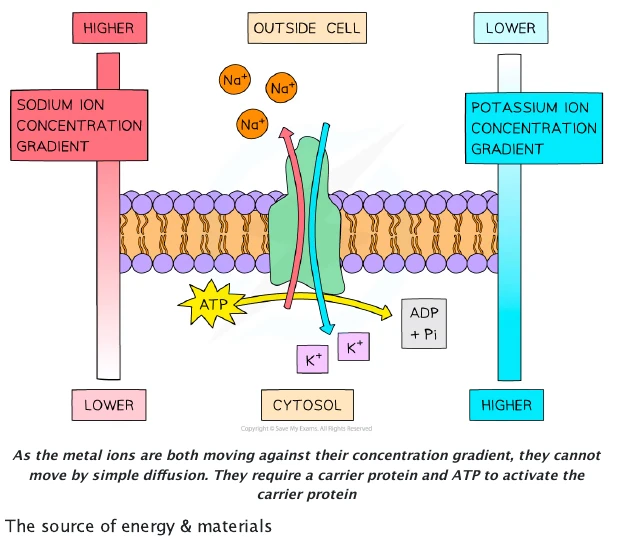
| Active transport | Against a gradient | Carrier/pump; ATP required |
| Co-transport | One solute down its gradient drives another | Carrier; gradient maintained by active transport |
Endocytosis and exocytosis move bulk material in vesicles and require ATP. They do not move substances directly through the phospholipid bilayer.
Diffusion and facilitated diffusion
Diffusion is the net movement of particles from a region of higher concentration to a region of lower concentration as a result of their random motion. Particles move in both directions, but more cross from high to low concentration until dynamic equilibrium is reached. At equilibrium particles still move, but there is no net movement.
The rate of diffusion increases with a steeper concentration gradient, greater exchange-surface area and higher temperature, and decreases with a longer diffusion distance. Smaller molecules generally diffuse faster. Across a membrane, lipid solubility and the number of suitable transport proteins also matter.
Simple diffusion through the phospholipid bilayer suits molecules such as oxygen and carbon dioxide. Facilitated diffusion is also passive, but uses hydrophilic channels or specific carriers for ions and polar substances. Channel proteins may be gated. A carrier binds its solute and changes shape; because carrier number is finite, transport can approach a maximum rate at high solute concentration.
Active transport and co-transport
Active transport moves a substance against its concentration or electrochemical gradient. A solute binds to a specific carrier, ATP is hydrolysed, the protein is phosphorylated and changes conformation, and the solute is released on the other side. The carrier then returns to its original state. Respiratory poisons or oxygen shortage reduce ATP supply and therefore reduce active transport.
In co-transport, movement of one ion down its electrochemical gradient supplies the energy to move another solute through the same carrier. The ion gradient must first be established by ATP-driven pumping, so co-transport is indirectly dependent on respiration. A carrier moving two substances in the same direction is a symporter; one moving them in opposite directions is an antiporter.
Endocytosis and exocytosis
Endocytosis brings material into a cell when the membrane surrounds it and pinches off as a vesicle. Phagocytosis takes in large solid particles, while pinocytosis takes in fluid and dissolved substances. Receptor-mediated endocytosis concentrates particular molecules after they bind to membrane receptors. In exocytosis, an internal vesicle fuses with the cell surface membrane and releases its contents, as in secretion of digestive enzymes or neurotransmitters.
Both processes require ATP for cytoskeletal movement, vesicle transport and membrane fusion. They also change membrane area, so endocytosis and exocytosis are balanced in cells with high rates of membrane turnover.
Osmosis and water potential
Osmosis is the net movement of water from higher water potential to lower water potential through a partially permeable membrane. Pure water has a water potential of zero; dissolved solute makes water potential more negative. Pressure potential can make total water potential less negative.
Solute potential, \(\Psi_s\), is zero for pure water and becomes more negative as solute concentration rises. Pressure potential, \(\Psi_p\), is normally positive in a turgid plant cell because the wall pushes back against the expanding protoplast. At equilibrium, water molecules still cross in both directions but there is no net movement because the water potentials are equal.
Osmosis in plant and animal cells
| External solution | Animal cell | Plant cell |
|---|---|---|
| Higher water potential than cell | Water enters; the cell swells and may lyse because there is no wall | Water enters; the vacuole expands and the cell becomes turgid as the wall resists expansion |
| Same water potential as cell | No net water movement; cell volume remains stable | No net water movement; the cell may be flaccid if pressure potential is low |
| Lower water potential than cell | Water leaves; the cell shrinks or becomes crenated | Water leaves; the cell becomes flaccid and may plasmolyse as the protoplast pulls away from the wall |
Incipient plasmolysis is the point at which the cell surface membrane has just begun to lose contact with the wall and pressure potential is approximately zero. The cell wall itself does not pull away; the protoplast, including its membrane, shrinks away from the wall.
Surface area to volume ratio
As a cell or organism grows, volume rises faster than surface area, so surface area to volume ratio decreases. There is then less exchange surface per unit of metabolically active tissue and diffusion distances are often longer. Small cells exchange substances rapidly; larger organisms require specialised exchange surfaces, ventilation and mass-transport systems.
Folded membranes, microvilli, flattened shapes and branching all increase surface area without an equivalent increase in volume. When comparing models, calculate both surface area and volume in consistent units before simplifying the ratio.
Investigating membrane permeability with beetroot
Beetroot vacuoles contain a red betalain pigment. Intact tonoplast and cell surface membranes retain it. Cut equal cylinders or discs, rinse cut surfaces until the wash water is clear, then expose samples for the same time to different temperatures or alcohol concentrations. Membrane disruption allows pigment to diffuse out.
Measure the surrounding solution's absorbance with a colorimeter. Control beetroot source, piece dimensions and surface area, solution volume and pH, exposure time, washing and mixing. Use a water blank, repeat each treatment and calculate a mean. Greater absorbance indicates more pigment leakage and therefore greater membrane permeability. At high temperature, phospholipids become more fluid and membrane proteins may denature; alcohol disrupts hydrophobic interactions in the bilayer.
Investigating diffusion
Visking tubing is a non-living model of a partially permeable membrane. If it contains starch and reducing sugar and is suspended in water, small sugar molecules can diffuse through its pores while larger starch molecules remain. Test samples from inside and outside with Benedict reagent and iodine, include initial controls, keep volumes and time constant, and recognise that Visking tubing lacks membrane proteins and does not model active transport.
Diffusion and surface area to volume ratio can be investigated with indicator agar. Cut geometrically similar cubes of different side length, place them in the same acid concentration and measure the time for colour change or the penetration distance after a fixed time. Smaller cubes have a larger surface area to volume ratio and a greater proportion of their volume is reached in the same interval. Use equal acid volume per cube, consistent mixing and accurate dimensions.
Estimating plant-tissue water potential
Cut equal potato cylinders, blot and record their initial mass, then place them in a range of known sucrose concentrations for the same time and temperature. Remove, blot in a standardised way and record final mass. Percentage change allows pieces with different starting masses to be compared.
Plot mean percentage change in mass against sucrose concentration. The x-intercept, where percentage change is zero, estimates the concentration with the same water potential as the tissue. Interpolate rather than selecting the nearest tested concentration. Repeat, calculate spread and control cylinder diameter and length, potato source, solution volume, immersion time, temperature and surface drying. The method estimates an average tissue value and assumes negligible solute entry and tissue metabolism during the experiment.
Active recall checkpoint
5The Mitotic Cell Cycle
Cell-cycle sequence
Cell grows and produces proteins and organelles.
DNA replicates; each chromosome becomes two sister chromatids.
Further growth and preparation for division.
Mitosis separates nuclei; cytokinesis divides cytoplasm.
DNA replication occurs during interphase, not prophase. Chromosome number is counted by centromeres: replication doubles DNA content but not chromosome number.
Chromosome structure and terminology
During most of interphase DNA is extended as chromatin, which allows genes to be transcribed. Before nuclear division it coils around histone proteins and supercoils into visible chromosomes. After S phase, each chromosome consists of two genetically identical sister chromatids joined at a centromere. The centromere contains a kinetochore where spindle microtubules attach.
Homologous chromosomes carry the same genes at the same loci but may carry different alleles; one homologue came from each parent. Sister chromatids are copied parts of one chromosome. A diploid nucleus contains two sets of chromosomes, whereas a haploid nucleus contains one. Mitosis separates sister chromatids and normally produces nuclei genetically identical to the parent nucleus.
Why the cell cycle is controlled
Progress through the cycle is regulated by checkpoints and changing activity of cyclins and cyclin-dependent kinases. The G1 checkpoint assesses cell size, nutrients, growth signals and DNA damage. The G2 checkpoint checks that replication is complete. The spindle checkpoint prevents anaphase until every chromosome is correctly attached to spindle microtubules from opposite poles.
A cell with severe damage may pause for repair, enter a non-dividing state or undergo apoptosis. These controls prevent damaged DNA being passed to daughter cells. The time spent in each phase varies with cell type: rapidly renewing epithelia divide often, while many differentiated cells remain outside the active cycle.
Mitosis
In prophase chromosomes condense and the spindle forms. At metaphase bivalents are not present; individual chromosomes align at the equator. During anaphase centromeres divide and sister chromatids move to opposite poles. Telophase restores nuclear envelopes and chromosomes decondense.
Mitosis preserves chromosome number and genetic information, enabling growth, tissue repair, asexual reproduction and replacement of cells.
| Stage | Events to recognise and describe |
|---|---|
| Prophase | Chromatin condenses into visible chromosomes; nucleolus disappears; centrosomes move apart and spindle develops; nuclear envelope breaks down later in the stage |
| Metaphase | Individual chromosomes align at the equator; each centromere is attached to spindle fibres leading to opposite poles |
| Anaphase | Centromeres divide; sister chromatids become daughter chromosomes and are pulled centromere-first to opposite poles |
| Telophase | Chromosomes reach the poles and decondense; spindle breaks down; nuclear envelopes and nucleoli re-form |
| Cytokinesis | An animal cell constricts with a cleavage furrow; plant Golgi vesicles fuse to form a cell plate and new wall |
Spindle microtubules shorten at the kinetochore and also move chromosome sets apart by interacting with microtubules from the opposite pole. Equal chromatid separation ensures each daughter nucleus receives one copy of every chromosome. Mitosis is nuclear division; cytokinesis is division of the cytoplasm and should not be used as a synonym.
Observing mitosis
Root tips are useful because their meristems contain many dividing cells. A preparation is softened, stained so chromosomes are visible, squashed into a thin layer and viewed systematically. Identify stages from chromosome behaviour rather than from a cell's approximate shape. Count cells in randomly selected fields or along a defined transect to reduce selection bias.
The proportion of cells observed in a stage estimates the proportion of cycle time spent there, provided cells enter the cycle asynchronously and the sample is representative. A high mitotic index may indicate rapid growth, tissue repair or a tumour, but does not by itself prove malignancy.
Telomeres and replicative lifespan
Telomeres are repetitive DNA sequences at the ends of linear chromosomes. They protect coding DNA from loss and prevent chromosome ends being recognised as broken DNA. Ordinary DNA polymerase cannot fully copy the end of the lagging strand, so telomeres become shorter after many divisions. When critically short, they can trigger senescence or apoptosis.
Telomerase extends telomeres using an internal RNA template. It is active in germ-line cells and many stem cells, but has low activity in most differentiated somatic cells. Reactivation in cancer cells helps them continue dividing; this does not initiate every cancer, but contributes to unlimited replicative potential.
Control, stem cells and cancer
Checkpoints prevent progression if DNA is damaged or spindle attachment is incomplete. Mutation of proto-oncogenes, tumour-suppressor genes or genes controlling apoptosis can produce uncontrolled division. A benign tumour remains local; a malignant tumour invades and may metastasise.
A stem cell is unspecialised, can divide repeatedly and can differentiate. Totipotent cells can form every cell type including extra-embryonic tissue; pluripotent cells can form almost all body cell types; multipotent adult stem cells form a narrower range. As cells differentiate, different sets of genes are expressed, so they develop specialised proteins, organelles and shapes despite having the same genome.
Stem cells can replace damaged tissue and provide models for testing drugs or studying development. Risks include immune rejection, infection, uncontrolled division and inappropriate differentiation. Embryonic sources raise ethical questions about embryo status and consent; induced pluripotent stem cells avoid embryo destruction but still require safety testing. A balanced evaluation must distinguish scientific risk from ethical judgement.
Mutations that activate proto-oncogenes can stimulate division, while mutations that inactivate tumour-suppressor genes remove restraints. Failure of DNA repair or apoptosis allows further mutations to accumulate. Benign tumours remain contained; malignant cells invade nearby tissues, enter blood or lymph, travel and form secondary tumours by metastasis. Carcinogens increase mutation probability rather than guaranteeing cancer.
Active recall checkpoint
6Nucleic Acids and Protein Synthesis
Nucleotide structure and DNA
A nucleotide contains a pentose sugar, phosphate and nitrogenous base. DNA nucleotides join by phosphodiester bonds to form antiparallel strands. Complementary base pairing is A-T and C-G; hydrogen bonding and base stacking stabilise the double helix.
RNA contains ribose and uracil and is usually single-stranded. mRNA carries a codon sequence, tRNA carries a specific amino acid and anticodon, and rRNA forms part of the ribosome.
Nucleotides and polynucleotides
A pentose sugar joined to a nitrogenous base is a nucleoside; addition of phosphate makes a nucleotide. Adenine and guanine are double-ring purines, while cytosine, thymine and uracil are single-ring pyrimidines. DNA contains deoxyribose and the bases A, T, C and G; RNA contains ribose and uses U in place of T.
A condensation reaction joins the phosphate on carbon 5 of one nucleotide to the hydroxyl group on carbon 3 of the next, producing a phosphodiester bond. Repetition creates a sugar-phosphate backbone with direction: one end is 5′ and the other 3′. A polynucleotide's base sequence carries information while its backbone provides structural continuity.
DNA double-helix structure
Two polynucleotide strands run antiparallel, one 5′ to 3′ and the other 3′ to 5′. Bases project inward and pair specifically: A with T using two hydrogen bonds, and C with G using three. A purine always pairs with a pyrimidine, keeping the helix width uniform. Many hydrogen bonds together stabilise the molecule, but each is weak enough to separate during replication and transcription.
Complementary base pairing means the sequence of one strand determines the other. If double-stranded DNA contains 24% adenine, it also contains 24% thymine; the remaining 52% is divided equally between cytosine and guanine. Percentages derived this way apply to the whole double-stranded molecule, not necessarily to one strand alone.
RNA structure and roles
| RNA | Structure | Role |
|---|---|---|
| mRNA | Single linear strand containing a sequence of codons | Carries a transcribed copy of genetic information from DNA to ribosomes |
| tRNA | Short strand folded by internal base pairing; has an anticodon and an amino-acid attachment site | Delivers a specific amino acid and pairs its anticodon with a complementary mRNA codon |
| rRNA | Combines with proteins to form small and large ribosomal subunits | Positions mRNA and tRNA and contributes to peptide-bond formation |
Semi-conservative replication
Helicase breaks hydrogen bonds and separates strands.
Each parental strand acts as a template.
DNA polymerase adds complementary nucleotides in the 5′ to 3′ direction.
Ligase seals fragments on the lagging strand.
Each daughter DNA molecule contains one parental and one newly synthesised strand.
Helicase unwinds the helix and breaks hydrogen bonds between complementary bases. Free activated DNA nucleotides align by complementary base pairing with each exposed template. DNA polymerase catalyses phosphodiester-bond formation and can extend a strand only at its 3′ end, so new DNA is synthesised 5′ to 3′.
At a replication fork, the leading strand can be made continuously towards the fork. The antiparallel template forces the lagging strand to be made discontinuously as Okazaki fragments, which DNA ligase joins. Each product contains one original strand and one newly synthesised strand, hence semi-conservative replication. Complementarity supports accuracy, while proofreading and repair reduce but do not eliminate errors.
From gene to polypeptide
During transcription, RNA polymerase uses the template DNA strand to make complementary pre-mRNA. Introns are removed and exons joined in eukaryotes. At a ribosome, tRNA anticodons pair with mRNA codons and peptide bonds join amino acids in the specified order.
The genetic code is triplet, degenerate and nearly universal. A substitution can be silent, missense or nonsense; an insertion or deletion may cause a frameshift.
Transcription
RNA polymerase binds near the start of a gene and locally separates the DNA strands. One DNA strand acts as the template. Complementary RNA nucleotides align, with U opposite A, and RNA polymerase joins them into an RNA strand. The RNA sequence matches the non-template DNA strand except that U replaces T. When the termination region is reached, the transcript is released and DNA re-forms its double helix.
In eukaryotes the first transcript can contain introns and exons. Splicing removes introns and joins exons to produce mature mRNA, which leaves through a nuclear pore. Alternative splicing can join exons in different combinations, allowing one gene to produce more than one polypeptide.
Translation
A ribosome binds mRNA and begins at a start codon. A tRNA carrying the corresponding amino acid binds when its anticodon is complementary and antiparallel to the codon. A second tRNA binds at the next codon, the ribosome catalyses a peptide bond, and it moves one codon along. Empty tRNA leaves while another charged tRNA arrives. At a stop codon no tRNA binds; the completed polypeptide is released and folds or is modified.
The genetic code is triplet because three bases specify one amino acid, non-overlapping because each base belongs to one codon, degenerate because most amino acids have more than one codon, and almost universal because the same codons specify the same amino acids in nearly all organisms. Degeneracy does not mean one codon can specify several amino acids.
Gene mutations and their consequences
| Mutation | Possible consequence |
|---|---|
| Base substitution | May be silent because of code degeneracy, missense if an amino acid changes, or nonsense if a stop codon forms |
| Base insertion or deletion | If not in multiples of three, shifts the reading frame and changes every downstream codon |
| Duplication or larger rearrangement | Can add amino acids, disrupt regulation or change gene dosage |
The effect depends on position and chemical consequence. A mutation in non-coding DNA may alter no expressed product or may affect a regulatory sequence. In coding DNA, a conservative amino-acid replacement may have little effect, whereas changing a residue in an active site, binding site or structural region can strongly alter function. Mutation creates new alleles randomly with respect to need.
Active recall checkpoint
7Transport in Plants
Transport tissues
Mature xylem vessels are dead, hollow and joined end to end. Lignin waterproofs and strengthens walls, bordered pits permit lateral movement and the absence of end walls lowers resistance. Phloem contains living sieve-tube elements supported by companion cells with many mitochondria and plasmodesmata.
Root hair cells provide a large surface area. Mineral ions may enter by active transport, lowering cell water potential so water enters by osmosis.
Recognising tissues in roots, stems and leaves
In a young dicot root, xylem forms a central star with phloem between its arms. This central position gives support against pulling forces and places transport tissue near the water-absorbing root surface. In a young dicot stem, vascular bundles form a ring: xylem lies towards the centre, phloem towards the outside and cambium between them. In a leaf vein, xylem is nearer the upper epidermis and phloem nearer the lower epidermis.
When interpreting a micrograph, use multiple features rather than colour. Xylem vessels have wide empty lumens and thick lignified walls. Sieve-tube elements have thinner walls and may show sieve plates; companion cells are smaller, contain nuclei and lie closely beside them. A plan diagram should show the distribution of tissues without drawing individual cells.
Xylem vessel elements
Immature vessel elements deposit lignin in rings, spirals or reticulate patterns. Their end walls and cell contents break down, producing a continuous low-resistance tube. Lignin prevents collapse under tension, waterproofs the wall and adds support. Annular and spiral thickening allow elongation in young organs, while more complete lignification gives mature tissue greater strength. Unlignified pits allow water to move between vessels and into surrounding cells, and provide a route around a blockage.
Phloem sieve tubes and companion cells
A mature sieve-tube element is living but has very little cytoplasm, no nucleus and few organelles, leaving space for translocation. End walls form sieve plates with pores through which phloem sap moves. Companion cells retain a nucleus, dense cytoplasm and many mitochondria; plasmodesmata connect them to sieve-tube elements. They supply ATP and proteins and carry out loading and unloading.
Transpiration stream
Water evaporates from mesophyll cell walls and diffuses through stomata, lowering leaf water potential. Water moves from xylem to mesophyll and tension pulls a continuous water column upward. Cohesion between water molecules and adhesion to xylem walls maintain the column.
Higher light intensity usually opens stomata; higher temperature increases evaporation; lower humidity steepens the water-vapour gradient; wind removes the humid boundary layer.
Water movement from soil to xylem
Mineral ions enter root hair cells through channel proteins or by active transport. This lowers cell water potential and water enters by osmosis. In the apoplast pathway, water moves through cell walls and intercellular spaces without crossing membranes. In the symplast pathway, water moves through cytoplasm and plasmodesmata. A vacuolar route additionally crosses tonoplasts.
The waterproof Casparian strip in endodermal cell walls blocks the apoplast. Water and ions must cross a cell surface membrane into the symplast, allowing selective transport proteins to control entry into the vascular tissue. Endodermal cells may actively move ions into xylem, lowering its water potential so water follows.
Cohesion-tension mechanism
Evaporation from moist mesophyll walls causes water menisci to curve and creates tension. Water moves from the xylem into the leaf down a water-potential gradient. Hydrogen bonding gives cohesion between water molecules, so tension is transmitted down an unbroken water column and pulls water upward as mass flow. Adhesion between water and hydrophilic xylem walls helps stabilise the column.
This pull is passive: xylem vessels do not use ATP because they are dead. Root pressure can make a small contribution when ions are actively accumulated in xylem, but it is not sufficient to explain water movement to the top of tall plants. Cavitation breaks a column; pits allow water to bypass the blocked vessel.
Measuring transpiration with a potometer
A bubble potometer estimates water uptake by following an air bubble in a capillary tube. Cut a leafy shoot under water to prevent air entering xylem, assemble the apparatus full of water, seal every joint, dry the leaves and check for leaks. After acclimatisation, record distance moved in a measured time. The reservoir can reset the bubble.
Change one environmental factor and control the others, including leaf area. Repeat with the same shoot where possible or normalise rate per unit leaf area. The method assumes water uptake approximates water loss, but some water is used in photosynthesis, growth and turgor maintenance.
Factors affecting transpiration
| Factor | Effect and explanation |
|---|---|
| Light intensity | Stomata usually open for photosynthesis, increasing the diffusion pathway for water vapour; response eventually plateaus when stomata are fully open |
| Temperature | Increases evaporation and molecular kinetic energy; very high temperature may close stomata if water stress develops |
| Humidity | High humidity reduces the water-vapour gradient between air spaces and atmosphere, lowering diffusion rate |
| Wind speed | Moderate wind removes the humid boundary layer and steepens the gradient; severe wind can trigger stomatal closure |
| Soil water | Low availability lowers leaf water potential and promotes stomatal closure, reducing transpiration |
Xerophytic adaptations
Xerophytes reduce water loss with a thick waxy cuticle, fewer stomata, stomata sunken in pits, rolled leaves and dense hairs. Pits, hairs and rolling trap still humid air, increasing boundary-layer resistance and reducing the diffusion gradient. Reduced leaf area lowers exchange surface. Succulent tissue stores water, while extensive shallow roots or deep roots improve uptake. Each adaptation must be linked to a specific change in gradient, distance, surface area or stomatal conductance.
Translocation and evidence
At a source, sucrose is actively loaded into companion cells and sieve tubes, reducing water potential. Water enters from xylem, raising hydrostatic pressure. At a sink sucrose is unloaded, water potential rises, and water returns to xylem. A pressure gradient drives mass flow.
Sucrose loading and mass flow
At a source such as a photosynthesising leaf, proton pumps use ATP to move \(\mathrm{H^+}\) out of companion cells. Protons then return through sucrose-proton co-transporters, carrying sucrose into the companion cell. Sucrose passes through plasmodesmata into a sieve-tube element, lowering its water potential. Water enters from adjacent xylem by osmosis and raises hydrostatic pressure.
At a sink such as a growing root, fruit or storage organ, sucrose leaves the sieve tube and is respired, converted to storage material or used in growth. Phloem water potential rises, water returns to xylem and hydrostatic pressure falls. The pressure difference between source and sink drives bulk flow. Different sieve tubes can transport in different directions at the same time, but sap in one tube has one net direction at a given moment.
Evidence for phloem transport
Removing a ring of bark removes phloem but leaves xylem. Sugars accumulate above the ring and tissues below receive less, supporting transport in phloem. Aphid stylets inserted into sieve tubes release sap under pressure; analysis reveals high sucrose and amino-acid concentrations. Radioactively labelled carbon dioxide supplied to a leaf appears first in sugars at the source and later at sinks, and autoradiographs trace movement. These observations support translocation, while metabolic inhibition reducing movement supports an energy-dependent loading step.
Active recall checkpoint
8Transport in Mammals
The heart and cardiac cycle
The sinoatrial node initiates excitation across the atria. A delay at the atrioventricular node allows ventricular filling; excitation then travels through the bundle of His and Purkyne tissue so ventricles contract from the apex upward.
During systole, rising ventricular pressure closes atrioventricular valves and later opens semilunar valves. During diastole, pressure falls, semilunar valves close and atrioventricular valves open. Valves respond to pressure differences rather than contracting.
Closed double circulation
Mammals have a closed circulation because blood remains in vessels, and a double circulation because it passes through the heart twice in one complete circuit. The pulmonary circuit sends deoxygenated blood from the right ventricle to lungs and returns oxygenated blood to the left atrium. The systemic circuit sends oxygenated blood from the left ventricle to body tissues and returns it to the right atrium.
Separation prevents oxygenated and deoxygenated blood mixing. High pressure can be generated for systemic flow, while pulmonary pressure remains lower to protect delicate lung capillaries. Continuous flow delivers oxygen and nutrients and removes carbon dioxide and other wastes more rapidly than diffusion alone could across a large body.
Heart structure
The venae cavae enter the right atrium; the pulmonary artery leaves the right ventricle. Pulmonary veins enter the left atrium; the aorta leaves the left ventricle. The atrioventricular valves lie between atria and ventricles, while semilunar valves lie at the bases of the pulmonary artery and aorta. Tendinous cords prevent atrioventricular valves inverting during ventricular systole.
The atria have thin walls because they move blood a short distance at low pressure. Ventricular walls are thicker; the left ventricle is thickest because it supplies the systemic circuit at high pressure. The septum prevents mixing. Coronary arteries branch from the aorta and supply myocardium with oxygen and glucose; blockage can cause myocardial infarction.
Pressure changes through one cardiac cycle
During atrial systole, atrial pressure rises and completes ventricular filling through open atrioventricular valves. Ventricular systole begins when ventricular pressure exceeds atrial pressure, closing these valves. There is briefly no change in ventricular volume while both valve sets are closed. When ventricular pressure exceeds arterial pressure, semilunar valves open and blood is ejected.
As ventricles relax, pressure falls below arterial pressure and semilunar valves close. When ventricular pressure drops below atrial pressure, atrioventricular valves open and passive filling begins. The sound “lub” is mainly associated with atrioventricular-valve closure and “dub” with semilunar-valve closure.
Coordination of the heartbeat
Cardiac muscle is myogenic, so it contracts without a motor nerve impulse. The sinoatrial node acts as pacemaker. Excitation spreads over atrial walls but is insulated from ventricles except at the atrioventricular node. The AVN delay allows the atria to finish emptying. The bundle of His conducts through the septum to Purkyne tissue, which spreads excitation from the apex upward so blood is pushed towards the arteries.
Autonomic nerves and hormones modify rather than initiate rhythm. Sympathetic stimulation and adrenaline increase heart rate, while parasympathetic stimulation decreases it. Stroke volume and heart rate determine cardiac output, which rises during exercise to meet greater oxygen demand and remove carbon dioxide.
Vessels and tissue fluid
| Vessel | Adaptation | Reason |
|---|---|---|
| Artery | Thick elastic and smooth-muscle wall; small lumen | Withstands and maintains high pulsatile pressure |
| Vein | Large lumen, valves, thinner wall | Returns blood at low pressure |
| Capillary | One-cell-thick endothelium; narrow lumen | Short diffusion distance and slow flow |
High hydrostatic pressure at the arteriole end forces plasma out, but cells and most proteins remain in blood. Lower pressure and the low water potential created by plasma proteins return fluid at the venule end; excess enters lymph vessels.
How vessel structure matches pressure and function
Arteries have collagen for strength, elastic fibres that stretch during systole and recoil during diastole, and smooth muscle that changes lumen diameter. Their folded endothelium permits expansion. Arterioles contain proportionally more smooth muscle and control distribution to tissues by vasoconstriction and vasodilation.
Veins carry blood at much lower pressure, so they have thinner walls and wide lumens that reduce resistance. Pocket valves prevent backflow, and contraction of surrounding skeletal muscle helps return blood. Capillaries consist of a single endothelial layer on a basement membrane. Their vast total cross-sectional area slows blood, and narrow lumens bring red cells close to the wall for exchange.
Blood cells and plasma
| Component | Key features and function |
|---|---|
| Red blood cell | Biconcave, flexible and without a nucleus or mitochondria; packed with haemoglobin for oxygen transport |
| Neutrophil | Multi-lobed nucleus and granular cytoplasm; engulfs pathogens by phagocytosis |
| Monocyte | Large cell with kidney-shaped nucleus; enters tissues and differentiates into a macrophage |
| Lymphocyte | Large spherical nucleus with a thin rim of cytoplasm; involved in specific immune responses |
| Platelet | Small cell fragment involved in clot formation |
| Plasma | Transports cells, nutrients, hormones, antibodies, carbon dioxide, urea, ions, proteins and heat |
Tissue fluid and lymph
At the arteriole end of a capillary, blood hydrostatic pressure exceeds the opposing effects of tissue-fluid pressure and the low water potential caused by plasma proteins. Water and small solutes leave through gaps between endothelial cells by ultrafiltration. Red cells and most plasma proteins remain, so tissue fluid resembles plasma but has far less protein.
Fluid bathes cells, allowing diffusion of oxygen and nutrients towards them and carbon dioxide and wastes away. Along the capillary, hydrostatic pressure falls while plasma proteins remain, making blood water potential lower. Most water therefore returns by osmosis near the venule end. Excess enters blind-ended lymphatic vessels and is ultimately returned to veins. Blocked lymph drainage can cause oedema.
Haemoglobin transport
Haemoglobin shows cooperative binding, producing a sigmoid oxygen-dissociation curve. At high partial pressure in lungs it loads oxygen; at lower partial pressure in respiring tissue it unloads. Carbon dioxide lowers pH and shifts the curve right (Bohr effect), promoting unloading.
Loading and unloading oxygen
Each haemoglobin molecule has four haem groups and can bind four oxygen molecules reversibly. The first oxygen is relatively difficult to bind, but its binding changes quaternary structure and increases affinity at remaining sites. This positive cooperativity creates the steep middle of the sigmoid curve, where a small fall in partial pressure produces substantial unloading.
In lung alveoli, high oxygen partial pressure promotes loading and haemoglobin becomes close to saturated. In actively respiring tissues, oxygen is consumed, partial pressure is lower and haemoglobin unloads. Fetal haemoglobin has a higher oxygen affinity than adult haemoglobin, helping transfer oxygen across the placenta.
Carbon dioxide transport and the Bohr effect
Some carbon dioxide dissolves in plasma and some binds to haemoglobin as carbaminohaemoglobin, but most enters red blood cells. Carbonic anhydrase catalyses its reaction with water to form carbonic acid, which dissociates into \(\mathrm{H^+}\) and \(\mathrm{HCO_3^-}\). Hydrogencarbonate diffuses into plasma while chloride ions enter to maintain electrical neutrality. Haemoglobin buffers hydrogen ions.
High carbon dioxide and hydrogen-ion concentrations reduce haemoglobin's oxygen affinity, shifting the dissociation curve to the right. This Bohr effect increases unloading in respiring tissues. In lungs, carbon dioxide is removed, pH rises and affinity increases, promoting loading. “Shift right” must be tied to lower affinity at a given oxygen partial pressure.
High-altitude responses
At altitude, lower atmospheric pressure lowers alveolar oxygen partial pressure and haemoglobin saturation. Immediate responses include faster ventilation and increased cardiac output. Over days to weeks, erythropoietin stimulates greater red-cell production, raising haemoglobin concentration. Capillary density and cellular adjustments may also increase. These changes improve delivery but very high haematocrit increases blood viscosity and workload on the heart.
Active recall checkpoint
9Gas Exchange
Exchange surface
Millions of alveoli provide a large surface area. Squamous epithelium and capillary endothelium give a short diffusion path, surfactant lowers surface tension, elastic fibres support recoil, and ventilation plus blood flow maintain steep oxygen and carbon-dioxide gradients.
Ventilation is bulk movement of air; gas exchange is diffusion across the alveolar-capillary barrier.
Organisation of the gas-exchange system
Air passes through the trachea, bronchi, bronchioles and alveolar ducts to alveoli. Trachea and bronchi are supported by cartilage, which prevents collapse while allowing flexibility. Smooth muscle changes airway diameter; elastic fibres help restore shape. Goblet cells secrete mucus that traps particles and pathogens, and ciliated epithelial cells move the mucus towards the throat.
Bronchi have cartilage plates, glands, smooth muscle and ciliated epithelium. Bronchioles have no cartilage and progressively less ciliated epithelium, but their smooth muscle can strongly regulate resistance. Alveoli have extremely thin walls and no cilia or cartilage. Recognise structures from combinations of lumen shape, cartilage, glands, epithelial type and surrounding alveoli.
Alveolar adaptations
| Feature | Contribution to rapid exchange |
|---|---|
| Very many alveoli | Create a huge total surface area |
| Squamous alveolar epithelium and capillary endothelium | Provide a diffusion pathway often only two thin cells plus basement membranes |
| Moist lining | Allows gases to dissolve before crossing membranes |
| Dense capillary network | Maintains blood flow and steep partial-pressure gradients |
| Ventilation | Continuously refreshes alveolar air |
| Elastic fibres | Allow stretch during inspiration and recoil during expiration |
| Surfactant | Reduces surface tension and helps prevent alveolar collapse, especially at low volume |
Gas exchange by diffusion
Alveolar oxygen partial pressure exceeds that in deoxygenated capillary blood, so oxygen dissolves in the moist lining and diffuses through epithelium, basement membranes and endothelium into plasma and red cells. Binding to haemoglobin helps keep dissolved oxygen concentration low and sustains the gradient. Carbon dioxide diffuses in the opposite direction because its partial pressure is higher in blood than alveolar air.
Large surface area, short distance and steep gradients all raise diffusion rate. Ventilation maintains alveolar gas composition while continuous perfusion brings deoxygenated blood and removes oxygenated blood. Fluid accumulation, fibrosis or destruction of alveolar walls lowers exchange by increasing distance or reducing surface area.
Ventilation mechanism
During inspiration the diaphragm contracts and flattens and external intercostal muscles lift the ribs. Thoracic volume increases, pressure falls below atmospheric pressure and air enters. Quiet expiration is mainly passive as muscles relax and elastic tissue recoils.
Pressure changes in ventilation
The pleural membranes surround the lungs with a thin lubricating fluid layer. Cohesion in this layer couples lung surface to the moving thoracic wall while reducing friction. During inspiration, external intercostal muscles contract, ribs move up and out, and the diaphragm flattens. Increased thoracic volume lowers intrapulmonary pressure below atmospheric pressure, so air flows inward down a pressure gradient.
During quiet expiration these muscles relax. Elastic recoil reduces thoracic and lung volume, raising pressure above atmospheric pressure and forcing air out. Forced expiration additionally uses internal intercostal and abdominal muscles. Air moves because of pressure differences; lungs are not filled because the diaphragm “pulls air” directly.
Interpreting lung-volume data
Tidal volume is the volume moved in one resting breath. Inspiratory and expiratory reserve volumes are the additional volumes that can be inhaled or exhaled beyond a normal breath. Vital capacity is the maximum volume exhaled after a maximal inhalation. Residual volume remains after maximal expiration and cannot be measured by a simple spirometer.
A spirometer trace can give breathing rate from cycles per minute and tidal volume from vertical change. If soda lime absorbs exhaled carbon dioxide and oxygen is supplied in a closed apparatus, a downward baseline trend represents oxygen uptake. Apply the apparatus calibration, state units and account for temperature and pressure where required.
Smoking and disease
Tar damages cilia and stimulates mucus production, increasing infection risk. Carcinogens raise mutation risk; elastase released during inflammation breaks down alveolar walls in emphysema. Carbon monoxide binds haemoglobin with high affinity and reduces oxygen transport.
Tar, mucus and chronic obstructive disease
Tar contains irritants and carcinogens. Irritation stimulates goblet cells and mucus glands to secrete more mucus, while damage to cilia reduces its removal. Mucus accumulates, obstructs airways and provides a site for pathogens, increasing infection and inflammation. Persistent inflammation can narrow bronchioles and cause chronic bronchitis, characterised by long-term productive cough.
Phagocytes entering inflamed lung tissue release elastase. Normally an inhibitor limits this enzyme, but cigarette smoke can reduce inhibitor effectiveness. Elastin and alveolar walls are destroyed, forming larger air spaces with less surface area and weaker recoil. In emphysema, expiration becomes difficult, air is trapped and oxygen diffusion falls.
Carcinogens, nicotine and carbon monoxide
Carcinogens increase mutation rate. If mutations activate proto-oncogenes, disable tumour-suppressor genes or impair DNA repair, cell division may become uncontrolled and lung cancer can develop. Cancer risk depends on dose, duration and other factors, so epidemiological evidence shows probability rather than certainty.
Nicotine binds receptors in the nervous system and promotes adrenaline release, increasing heart rate and blood pressure. It is addictive and can cause vasoconstriction, adding cardiovascular strain. Carbon monoxide binds haemoglobin far more strongly than oxygen, forming carboxyhaemoglobin. This reduces oxygen-carrying capacity and makes remaining haem groups hold oxygen more tightly, reducing unloading to tissues.
Evaluating smoking evidence
Look for dose-response relationships, large representative samples, consistent findings and plausible biological mechanisms. Control or discuss confounders such as age, occupation, pollution and socioeconomic factors. Relative risk compares proportions between groups, but absolute risk is needed to judge the number of people affected. An observational association cannot alone prove causation, yet converging epidemiological and mechanistic evidence can make a causal conclusion strong.
Active recall checkpoint
10Infectious Diseases
Transmission and prevention
A pathogen causes disease; a vector transfers a pathogen between hosts. Transmission may be direct, through droplets, contaminated food or water, body fluids, surfaces or vectors. Prevention must interrupt the relevant route.
Community control combines clean water, sanitation, food hygiene, ventilation, barrier protection, vector control, vaccination, contact tracing and isolation where proportionate.
Pathogen, infection and disease
A pathogen is an organism or acellular agent capable of causing disease. Infection occurs when it enters and multiplies in a host; disease refers to impaired normal functioning and symptoms. A person can be infected without obvious symptoms and still transmit a pathogen. A reservoir is the usual habitat in which a pathogen persists, while a vector is a living organism that transfers it between hosts.
Direct transmission includes physical or sexual contact and short-range droplets. Indirect transmission includes contaminated food, water, objects, blood products, airborne particles and vectors. The route determines effective control: water treatment cannot by itself prevent a mosquito-borne infection, and antibiotics cannot interrupt viral replication.
Key CAIE disease examples
| Disease and pathogen | Transmission and important biology | Control |
|---|---|---|
| Cholera - Vibrio cholerae bacterium | Faecal contamination of water or food; toxin causes chloride secretion from intestinal epithelial cells, so water follows into the lumen and severe watery diarrhoea develops | Clean water, sanitation, hand and food hygiene, vaccination in risk settings; oral rehydration replaces water and ions |
| Malaria - Plasmodium protoctist | Female Anopheles mosquito vector; stages reproduce in liver and red blood cells, whose rupture contributes to periodic fever and anaemia | Insecticide-treated nets, indoor spraying, drainage of breeding water, larval control, antimalarial drugs and rapid diagnosis |
| Tuberculosis - Mycobacterium tuberculosis | Airborne droplets; bacteria survive in lung tissue and may remain latent before active disease | Ventilation, detection and contact tracing, BCG where recommended, and a full combination-drug course |
| HIV/AIDS - human immunodeficiency virus | Sexual transmission, infected blood or shared needles, and parent-to-child transmission; infects helper T cells and progressively weakens specific immunity | Barrier protection, screened blood, sterile needles, testing, antiretroviral therapy and prevention of mother-to-child transmission |
Why disease patterns differ
Transmission depends on climate, vector range, population density, housing, sanitation, access to clean water, vaccination, health services, travel, conflict and public trust. Poverty can increase exposure and reduce access to diagnosis and treatment. Climate change can alter vector distribution, but local ecology and control measures determine whether transmission actually increases.
Endemic means a disease is consistently present in a region; an outbreak is a rise above the expected level; an epidemic affects many people in a community or region; a pandemic is sustained across countries or continents. These terms describe distribution, not how severe an individual case is.
Treatment and resistance
Antibiotics target bacterial structures or processes and do not treat viruses. Random mutation creates variation; antibiotic exposure is a selection pressure, susceptible bacteria die, resistant bacteria survive and reproduce, and resistance alleles spread vertically or through horizontal gene transfer.
How penicillin works
Penicillin is a beta-lactam antibiotic. It binds to bacterial enzymes that cross-link peptidoglycan during cell-wall synthesis. Growing bacteria then form weakened walls and may burst as water enters. Human cells lack peptidoglycan, giving selective toxicity. Penicillin does not kill viruses, which have no bacterial wall and replicate using host-cell machinery.
Some bacteria produce beta-lactamase, which breaks the antibiotic's beta-lactam ring; others alter the target protein, reduce entry or pump the drug out. Resistance does not mean the human body has become resistant. The resistant bacterial population has become more frequent.
Evolution and spread of antibiotic resistance
Mutation or acquisition of a resistance gene creates variation before treatment. Antibiotic exposure kills susceptible cells, leaving resistant cells with less competition. They reproduce by binary fission and pass resistance vertically. Plasmids can also transfer between bacteria by conjugation, and DNA can be exchanged by transformation or bacteriophage-mediated transfer.
Unnecessary prescribing, use against viral disease, incorrect doses, incomplete courses, unregulated access and routine use in livestock all increase selection. Resistance genes and resistant strains can spread through hospitals, travel, food systems, wastewater and poor infection control. Multi-drug resistance makes treatment longer, more toxic and more expensive and increases mortality.
Control combines antibiotic stewardship, susceptibility testing, narrow-spectrum treatment where suitable, correct dosing, patient adherence, vaccination, hygiene, surveillance and development of new treatments. Reducing antibiotic use does not instantly remove all resistance because genes may carry little fitness cost or remain linked to other selected genes.
Evaluating epidemiological evidence
Compare incidence or prevalence between groups, consider sample size, confounders, diagnostic criteria and time period, and quote data with units. A controlled trial tests an intervention more directly than an observational correlation.
Prevalence counts existing cases; incidence counts new cases arising in a defined population over time.
Incidence, prevalence and mortality
Incidence measures risk of developing disease; prevalence reflects both incidence and how long people remain affected. Effective treatment that prolongs life can increase prevalence even while incidence falls. Mortality rate counts deaths in a population, while case-fatality proportion counts the fraction of diagnosed cases who die.
Designing and judging intervention studies
Define the population, outcome and follow-up period. Compare like groups, use random allocation where ethical, blind participants or assessors when possible and include a suitable control. Large sample size reduces random sampling error, while repeats across settings test generalisability. Report absolute numbers as well as percentages and include uncertainty such as confidence intervals.
Confounding occurs when another variable is associated with both exposure and outcome. Bias can arise from non-random recruitment, loss to follow-up, inaccurate self-reporting or changing diagnostic definitions. A statistically significant association may be small or not biologically important, and lack of significance may reflect insufficient power.
Active recall checkpoint
11Immunity
Non-specific and specific defence
Skin, mucus, cilia and stomach acid reduce entry. Phagocytes recognise, engulf and digest pathogens, then macrophages may present antigens. Specific responses are directed against antigens and generate memory.
A B lymphocyte with a complementary receptor undergoes clonal selection. Plasma cells secrete antibodies; memory B cells persist. Helper T cells release cytokines, cytotoxic T cells kill infected cells, and memory T cells accelerate later responses.
Phagocytosis
Receptors on a neutrophil or macrophage bind molecules on a pathogen or antibodies attached to it.
The membrane surrounds the pathogen and encloses it in a phagosome.
Lysosomes fuse with the phagosome and hydrolytic enzymes break down the pathogen.
A macrophage can display pathogen-derived antigen on its surface for recognition by helper T cells.
Phagocytosis is non-specific because the same cellular process acts against many pathogens. Antigen presentation links it to the specific response. Neutrophils are short-lived and abundant in blood; macrophages arise from monocytes, live longer in tissues and are important antigen-presenting cells.
Antigens and clonal selection
An antigen is a molecule recognised as foreign and capable of stimulating a specific immune response. It often occurs on a pathogen surface, but toxins, transplanted cells and altered body cells may also bear antigens. Each lymphocyte clone carries receptors with one binding-site shape. Only a lymphocyte whose receptor is complementary to the antigen is selected.
After activation and helper-T-cell signalling, the selected cell divides by mitosis. This clonal expansion produces many genetically identical cells with the same specificity. Effector cells act immediately, while memory cells remain and provide long-term capacity for a more rapid response.
Humoral and cell-mediated responses
| Cell | Main role |
|---|---|
| Helper T cell | Recognises presented antigen and secretes cytokines that stimulate specific B cells, cytotoxic T cells and phagocytes |
| Cytotoxic T cell | Recognises antigen on infected or abnormal body cells and releases molecules that trigger cell death |
| B lymphocyte | Binds intact complementary antigen, internalises it and can present it to helper T cells |
| Plasma cell | Has abundant RER and Golgi and secretes large quantities of one specific antibody |
| Memory B and T cells | Persist after the primary response and respond rapidly on re-exposure |
Humoral immunity is mediated by antibodies in body fluids and is particularly effective against extracellular pathogens and toxins. Cell-mediated immunity uses T cells against infected or abnormal body cells. The responses cooperate rather than occurring as isolated systems.
Primary and secondary immune responses
During first exposure there is a lag while a rare complementary lymphocyte is selected and clonally expanded. Antibody concentration rises, then falls as antigen is removed and many effector cells die. Memory cells remain. On later exposure to the same antigen, they divide quickly, producing a response with a shorter lag, a faster rate and a higher, longer-lasting antibody concentration. Disease may be prevented because the pathogen is removed before symptoms develop.
Autoimmune disease: myasthenia gravis
In an autoimmune disease, the immune system responds against a self antigen. In myasthenia gravis, antibodies bind to or promote destruction of acetylcholine receptors at neuromuscular junctions. Fewer functional receptors mean end-plate depolarisation is less likely to reach threshold, producing fatigable muscle weakness. Treatment may increase acetylcholine availability, suppress immune activity or reduce production of abnormal antibodies.
Antibodies and vaccination
Each antibody has variable regions forming specific binding sites and constant regions that interact with immune cells. Antibodies neutralise toxins or viruses, agglutinate cells and promote phagocytosis.
Vaccination introduces antigen safely, producing primary response and memory. A later exposure produces a faster, larger secondary response before disease develops. Herd protection reduces transmission when enough susceptible contacts are removed.
Antibody structure and action
An antibody is a globular protein made of two identical heavy chains and two identical light chains held by disulfide bonds. The tips contain variable regions; one variable region from a heavy and light chain together forms a binding site complementary to one epitope. The hinge gives flexibility. The constant region determines interactions with receptors on immune cells.
| Action | How it protects |
|---|---|
| Neutralisation | Binding blocks a toxin's active region or prevents a virus attaching to host receptors |
| Agglutination | Each antibody binds more than one particle, forming clumps that are easier for phagocytes to remove |
| Opsonisation | Antibody-coated pathogens bind more readily to phagocyte receptors |
| Complement activation | Constant regions help trigger a protein cascade that promotes inflammation and membrane damage |
Active and passive immunity
| Type | Source and consequence |
|---|---|
| Natural active | Infection exposes antigens; the person's own lymphocytes respond and make memory cells |
| Artificial active | Vaccination supplies antigen safely; the person's own response produces memory |
| Natural passive | Ready-made maternal antibodies cross the placenta or enter in breast milk; protection is immediate but temporary |
| Artificial passive | Injected antibodies give rapid short-term protection or treatment, but do not create memory cells |
Active immunity has a slower onset but can be long-lasting because memory forms. Passive immunity acts immediately but declines as transferred antibodies are broken down. Receiving antibody is not vaccination unless an antigen is also used to activate the recipient's own lymphocytes.
How vaccines control disease
Vaccines may contain killed pathogens, attenuated pathogens, purified antigens, toxoids or genetic instructions that cause host cells to make an antigen. The antigen cannot cause the full disease in an immunocompetent person but stimulates clonal selection, plasma-cell formation and memory. Booster doses restore or broaden memory where the first response declines.
High coverage reduces the number of susceptible hosts and breaks chains of transmission, indirectly protecting people who cannot be immunised or do not respond. The coverage needed depends on transmissibility and vaccine effectiveness. Antigenic change, poor uptake, cold-chain failure, unequal access and short-lived immunity can prevent elimination. Herd protection applies only to infections transmitted between people, not to a non-communicable disease.
Monoclonal antibodies
A selected B lymphocyte can be fused with a myeloma cell to create a hybridoma that both secretes one antibody and divides indefinitely. Monoclonal antibodies are used in tests, imaging and targeted treatment.
Producing monoclonal antibodies
Expose an animal to the target antigen so complementary B lymphocytes clonally expand.
Remove spleen B cells and fuse them with dividing myeloma cells to form hybridomas.
Use selective medium so unfused cells do not persist; screen supernatants for the required antibody.
Clone one positive hybridoma and culture it at scale; collect and purify its identical antibodies.
A B cell supplies the required antibody gene expression but does not divide indefinitely; a myeloma cell divides indefinitely but is selected not to produce its own antibody. The hybridoma combines both useful properties. “Monoclonal” means all antibodies originate from one cell clone and bind the same epitope.
Uses and evaluation of monoclonal antibodies
Diagnostic tests use a labelled monoclonal antibody to reveal a target such as a hormone, pathogen antigen or biomarker. Pregnancy tests use antibodies against hCG in a lateral-flow format, with a separate control line confirming that liquid has moved correctly. In imaging, an antibody can carry a fluorescent or radioactive label to locate particular cells.
Treatment can use an antibody to block a receptor, neutralise a signalling molecule, mark a target for immune destruction or deliver a drug or radioisotope to cells with a particular antigen. High specificity can reduce exposure of healthy cells, but the target may also occur on healthy tissue, tumours may vary or mutate, and antibodies can provoke immune reactions. Benefits, false results, cost and animal-use ethics must be evaluated for the stated application.